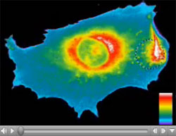
 NIH is seeking broad input from the scientific community on challenges and opportunities in single-cell analysis, a topic of great interest and relevance to many NIGMS grantees and applicants. Please help NIH shape its future programs in this emerging research area by sending in your opinions. The request for information (RFI) asks for responses on topics including:
- Current conceptual, technical and/or methodological challenges in the field;
- Major biomedical research questions that can be addressed by single-cell analysis; and
- The highest priority tools and resources needed to move forward.
We hope you’ll take the time to weigh in with your opinions and specific examples between now and the March 18 response deadline.
Let me know if you would like to learn more about trans-NIH activities in this area, as I’m a member of the group that issued this RFI—the Single Cell Analysis Working Group of the NIH Common Fund (formerly known as the NIH Roadmap), which provides strategic planning, coordination and support for programs that cut across NIH institutes and centers.


In our experience, access to sensitive, quantitative microscopy is a significant bottleneck. Most of these single cell assays use FRET as the readout. Going in vivo with FRET poses significant challenges. Measurements of fluorescence lifetime is the optimal method but sensitivity is low. Access to lifetime instruments with sensitivity that is comparable to intensity measurements would greatly facilitate work in this area.
Our work has been strengthened by methods that we developed to count fluorescent fusion proteins in live fission yeast cells (Wu, J.-Q. and Pollard, T.D. (2005) Counting cytokinesis proteins globally and locally in fission yeast. Science 310:310-314.) Coding sequences for fluorescent proteins are integrated into the genome, so each copy of the protein of interest is tagged and expressed from the native promoter. This allows for important controls to verify the functionality of the fusion protein.
Being able to count the number of fusion proteins in each pixel of a fluorescence micrograph over time in a live cell has allowed us to make the quantitative measurements required to formulate and test mathematical models for cytokinesis (Vavylonis, D., Wu, J.-Q., Hao, S., O’Shaughnessy, B. and Pollard, T.D. (2008) Assembly mechanism of the contractile ring for cytokinesis by fission yeast. Science 319:97-100.) and clathrin-mediated endocytosis (Berro, J., Sirotkin, V. and Pollard, T.D. (2010) Mathematical modeling of endocytic actin patch kinetics in fission yeast: disassembly requires release of actin filament fragments. Molec. Biol. Cell 21:2905-2915.).
All fields of cell biology would benefit from this approach, especially if barriers to integrating the coding sequences of the fluorescent proteins into the genomes could be overcome. The rest of the work can be done with standard fluorescence microscopes (at least after they have been calibrated).
Single cell analysis has historically provided enormously detailed data sets. On of the most striking example is the measurement of ion channel current in single cells and single channels. However, many more single cell techniques have since led to a much better understanding of cellular function.
The main challenges are (i) the development of high throughput adaptations of such single cell studies and (ii) implementation of a data base where single cell data is deposited making such data accessible in a way similar to sequence or structural information of genes and proteins.
Technologies that allow detection of specific protein-protein interactions in primary, unmodified (read: non-transfected) cells would help translate the wealth of information gleaned from FRET/FLIM experiments into an understanding of how such interactions occur, say, in patient-derived cells. Also, there is a growing need for methods that allow single cells to be analyzed within multidimensional environments that not only approximate the normal physiological environment of the cell in question but also remain experimentally manipulable and tractable.